

Home » Course Layouts » Free Course Layout Udemy
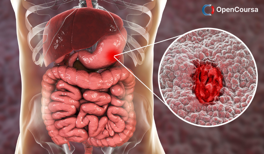
Histology is a key part of modern medicine – it helps us study cells and tissues at a microscopic level, and accurately diagnose diseases such as cancer.
0
52
English
English [CC]

- Learn basic syntax that can apply to any language.
- Learn what is a programming language and the basic concepts for beginners.
- Understand what is Javascript in it's truest form.
- Know the basic syntax of Javascript.
- Know some hidden quirks in Javascript.
Description
This course, Histology, microscopy, anatomy and disease, will help you understand the basic principles of light microscopy, before introducing you to histology, concentrating on the structure, function and relationship of normal human tissues.
As part of the course you will use a virtual microscope, enabling you to compare normal tissues with pathological (or diseased) tissues, and find out how pathologists use histopathology to examine tissues at a microscopic level and identify disease.
You will be making extensive use of the virtual microscope. It will work on all modern browsers on both desktops and tablets. However, it is recommended that you complete the course on a desktop for better viewing of the sections and to enable integration of microscopy with the course text, images and videos in separate windows.
This course is designed for students studying human biology at school or university, medical laboratory scientists and anyone who is interested in biomedical science.
By enrolling on this course you can track your progress in your My OpenLearn profile and earn a free Open University statement of participation on completion.
This course is also available to study on FutureLearn where you will have the opportunity to purchase a FutureLearn certification on completion.
This OpenLearn science course is produced with the kind support of Dangoor Education, the educational arm of The Exilarch's Foundation.
The Open University would really appreciate a few minutes of your time to tell us about yourself and your expectations of the course. We welcome your feedback and suggestions to improve the experience for other learners.
Course learning outcomes
After studying this course, you should be able to:
- Understand the basic principles of microscopy
- Identify a number of the more common tissues from their histological appearance
- Relate the structure of tissues seen under the microscope to their function
- Identify gross abnormalities, such as neurodegeneration, inflammation and cancer.
Course content
-
- Introduction 00:15:00
-
- What is histology? 00:20:00
- A histology department in action 00:25:00
- How histological slides are produced 00:20:00
- Staining techniques 00:25:00
- Immunohistochemistry 00:15:00
- Cells and tissues 00:20:00
- Using a light microscope 00:20:00
- Basic functions of the virtual microscope 00:10:00
- Normal blood smear 00:15:00
- Malaria 00:15:00
- Considerations for interpreting sections 00:20:00
- Introduction 00:05:00
- Identifying tissues 00:10:00
- Introduction to different tissues 00:20:00
- Structure of a lymph node 00:20:00
- Immunohistochemistry 00:20:00
- Immunohistochemistry technique 00:10:00
- Using IHC to identify B cells and T cells 00:10:00
- Putting it into practice 00:15:00
- Summary of Week 2 00:10:00
- Functions of tissues 00:25:00
- Structure–function relationships 00:15:00
- Introduction 00:10:00
- Cell death and degeneration 00:30:00
- Cell death in the CNS 00:20:00
- Histopathology of the nervous system 00:15:00
N.A
- 5 stars0
- 4 stars0
- 3 stars0
- 2 stars0
- 1 stars0
No Reviews found for this course.